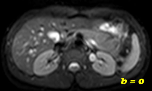
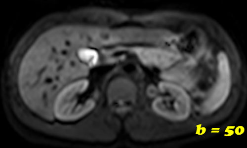
A baseline b-value of 50 s/mm² is often used in liver diffusion-weighted imaging instead of b = 0. The reason is readily apparent from the images below.
With b = 0 bright signals are noted in multiple veins due to the high T2 of blood coupled with sluggish flow. This array of multiple white dots on the b0 image makes distinguishing cysts, masses, and vessels difficult. By raising the starting b-value to a low number like 50-100, these vascular white dots disappear, providing a cleaner visualization of the hepatic parenchyma.
With b = 0 bright signals are noted in multiple veins due to the high T2 of blood coupled with sluggish flow. This array of multiple white dots on the b0 image makes distinguishing cysts, masses, and vessels difficult. By raising the starting b-value to a low number like 50-100, these vascular white dots disappear, providing a cleaner visualization of the hepatic parenchyma.
The reason the vascular dots disappear with low b-values is that flowing protons within them have moved to a different position in the gradient field. Because of motion these protons will not regain their original phase information when the diffusion rephasing gradient is applied and will undergo signal loss.
Advanced Discussion (show/hide)»
Other than liver imaging, I know of no reason why body imagers would choose b=50 instead of b=0 starting values elsewhere in the abdomen and pelvis. Perhaps it is simply habit that b=50 values are used in these other areas.
Unless multi-component ADC values need to be calculated, this small difference in starting b-values (0 vs 50) makes no significant difference in the single-component ADC maps commonly used.
References
Qayyum A. Diffusion-weighted imaging in the abdomen and pelvis: concepts and applications. Radiographics 2009; 29:1797-1810.
Qayyum A. Diffusion-weighted imaging in the abdomen and pelvis: concepts and applications. Radiographics 2009; 29:1797-1810.
Related Questions
What is meant by the b-value? How do I pick it?
What is meant by the b-value? How do I pick it?


