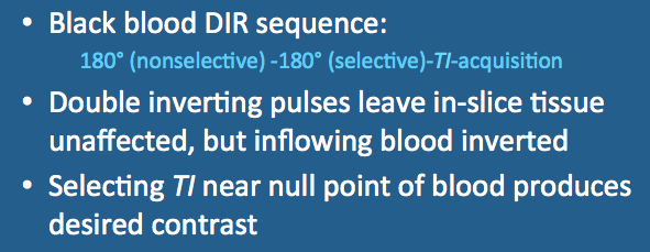
Over the next several hundred msec, two events occur simultaneously: 1) inverted blood initially outside the imaged slice undergoes T1-recovery of its longitudinal magnetization, passing through zero as it attempts to realign with Bo; and 2) this outside blood flows into and replaces blood within the imaged slice.
The inversion time (TI) is chosen so that the magnetization of this inflowing blood passes through zero at the start of image acquisition. For cardiac imaging at 1.5T with a heart rate of 60 BPM, this typically means a TInull of approximately 650 msec. If the HR is too fast, TI must be reduced. Some systems do this automatically using a software-based blood suppression TI calculation.
The most commonly used imaging sequence in conjunction with this double IR method is fast (turbo) spin echo with a relatively short TEeff (≈ 40 msec). Alternatively, a segmented k-space gradient echo or echo-planar sequence may also be used.
First relatively long inversion times are necessary especially at slower heart rates to achieve sufficient nulling of blood. These long TI values may conflict with optimal trigger delay, forcing adoption of a suboptimal imaging protocol. With long TI's a repetition time (TR) of two heart beats may be required for effective blood suppression. The end result is that imaging of a single coronary artery might take in excess of 10 minutes to image using the DIR method.
|
Blood suppression may also be compromised if the plane of imaging is not perpendicular to the direction of flow. This is illustrated the image (right) where signal in the cardiac chambers is well-suppressed but that in the descending aorta (red arrow) is not. Here flow in the aorta lies within the plane of imaging, and aortic blood is is not completely replenished between the two inversion pulses. A similar phenomenon may occur in tortuous or slowly-flowing arteries. Sometimes an artifactual high-intensity band may appear along the periphery of an artery due to slow laminar flow at the boundary between the vessel wall and its lumen.
|
Advanced Discussion (show/hide)»
Choice of Image Acquisition Module for DIR Imaging
Several MR sequences are available to produce dark blood imaging. These include fast (turbo) SE, HASTE, TrueFISP, and FLASH. Depending on the technique, T1- and/or T2-weighted images may be produced. As blood will be always be dark by definition, T1- and T2-weighting refers to image contrast in the non-suppressed solid tissues (myocardium, chest wall, etc.)
The usual purpose for dark-blood DIR imaging is to assess cardiac anatomy, and hence weighting is not particularly important. To minimize imaging time, TR is chosen to be as short as possible, resulting in T1-weighted images. For certain applications, however, such as evaluation of cardiac masses, T2-weighted imaging may be preferred.
The most common image acquisition module used for DIR dark blood imaging is a turbo (fast) spin echo technique. This is typically performed in segmented, single-slice mode with breath-holding or a navigator. T1-weighted contrast is achieved using a short TR (triggering on every heart beat) and short TEeff (10-30 msec, requiring a low turbo factor). Alternatively, T2-weighted images may be obtained by using a long TR (triggering on every other heart beat) and a longTEeff.
The second common method for dark blood DIR uses HASTE/single-shot variant of fast (turbo) SE. This is ideally performed at one slice per heartbeat with breath-holding, but can be done with free breathing in uncooperative patients. Because the long echo train used in HASTE means a long effective TE (>100 ms), HASTE dark blood sequences are by necessity T2-weighted.
Gradient echo techniques may also be used for dark blood cardiac MRI, but these are less common. Segmented or single-shot TrueFISP methods produce T2/T1 contrast, while FLASH/SPGR methods create T1-weighted images.
Additional Comments about DIR Parameters and Pulse Sequence Design
As described in a prior Q&A, in a prospectively gated sequence TR cannot be independently prescribed but depends on the heart rate (HR). A faster HR translates to a shorter RR interval and less recovery time (TR) for the sequence. Accordingly, the dark blood inversion time (TI) is shorter at faster HR's. For example, at a HR of 60 BPM, the TR is 2000 ms and the optimal dark blood TI at 1.5T is about 630 ms. At a HR of 100 BMP, the TR is 1200 and the optimal TI is about 420 ms.
Dark blood parameters must be specified in setup. This includes the relative thickness of the dark blood slab as well as the flip angle. Typically the DB slab inversion thickness is taken to be about 2x the actual slice thickness. To insure complete inversion of the blood signal across the entire slab, the flip angle of the second inverting pulse is often chosen to be slightly greater than 180° (e.g., 200° is typical).
The initial nonselective 180°-inversion pulse may be either a simple hard pulse or an adiabatic inversion pulse. The selective 180°-pulse is usually either adiabatic hyperbolic secant pulse or nonadiabatic Shinnar-Le Roux pulse. Spoiler gradients are often applied after the second inversion pulse to disperse any residual transverse magnetization.
Edelman RR, Chien D, Kim D. Fast selective black blood MR imaging. Radiology 1991; 181:655-660.
Liu Y, Riederer SJ, Ehman RL. Magnetization-prepared cardiac imaging using gradient echo acquisition. Magn Reson Med 1993; 30:271-275.
Siemens. Cardiac MRI Morphology 2004. (Slide-based review of dark blood technique with optimization suggestions and imaging examples).
Why go to the trouble of using a double IR method to suppress blood? Couldn't blood be suppressed with a single carefully selected TI value?
Isn't double inversion recovery enough? Why would you want to do triple IR?


