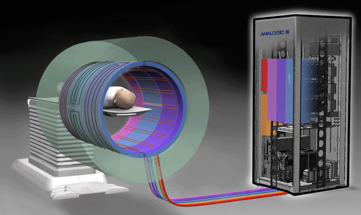
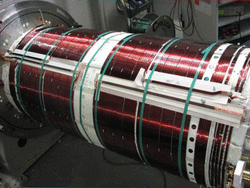
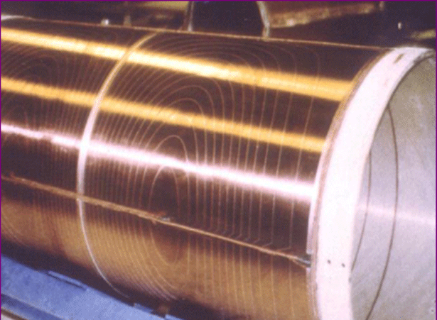
Historically, gradients coils were were composed of individual wires wrapped onto cylindrical formers made of fiberglass and coated with epoxy resin. Many laboratory instruments and very-high-field human scanners still use this method. Today, however, most widely manufactured superconducting scanners utilize distributed windings in a "fingerprint" pattern consisting of multiple thin metallic strips or large copper sheets etched into complex patterns and applied to the cylinder.
|
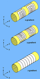
Three sets of gradient coils are used in nearly all MR systems: the x-, y-, and z-gradients. Each coil set is driven by an independent power amplifier and creates a gradient field whose z-component varies linearly along the x-, y-, and z-directions, respectively. The design of the z-gradients is usually based on circular (Maxwell) coils, while the transverse (x- and y-) gradients typically have a saddle (Golay) coil configuration. The physical principles underlying these designs will be considered in subsequent questions.
|
Applying a gradient causes a frequency variation of protons as a function of position along the direction of the gradient. This change in frequency can be used for spatial encoding. If the gradient is played out during slice selection and again during signal readout, a slice can be selected perpendicular to the gradient direction. For example, if the z-gradient is turned on in this fashion, a transverse slice is created in a supine patient. Oblique slices can be obtained by turning on two or more gradients simultaneously.
|
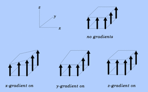
A frequent misconception about gradient fields is that the x- and y-gradients somehow skew or shear the main (Bo) field transversely. That is not the case as is shown in the diagram to the right. The x- and y-gradients provide augmentation in the z-direction to the Bo field as a function of left-right or anterior-posterior location in the gantry. The x- and y-gradients (ideally, at least) do not produce substantial components within the bore perpendicular to Bo.
|
Advanced Discussion (show/hide)»
No supplementary material yet. Check back soon!
References
Hidalgo-Tabon SS. Theory of gradient coil design methods for magnetic resonance imaging. Concepts Mag Res Part A 2001; 36A:223-242.
Lauterbur PC. Image formation by induced local interactions: examples employing nuclear magnetic resonance (pdf). Nature 1973; 242:190-191. (The classic paper demonstrating how gradients can be used for imaging that won Lauterbur the Nobel Prize in 2003).
Related Questions
What is a gradient?
I'm confused. Isn't a gradient some type of coil?
How do you make a z-direction gradient?
How do you make x- and y-direction gradients?
All the diagrams I have seen for gradients deal with cylindrical scanners. How are the gradients designed for open-bore scanners?
What is a gradient?
I'm confused. Isn't a gradient some type of coil?
How do you make a z-direction gradient?
How do you make x- and y-direction gradients?
All the diagrams I have seen for gradients deal with cylindrical scanners. How are the gradients designed for open-bore scanners?

