|
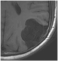
To the right are images of a brain tumor with intrinsically long T1 and T2 values having opposite intensities on T1- and T2-weighted images. To understand this "paradox", you must realize that a pixel's "brightness" or "darkness" on an MR image is directly related to the magnitude of the detected MR signal. The magnitude of the MR signal after an RF-pulse is in turn, dependent on two factors:
|
|
|
T1 reflects the length of time it takes for regrowth of Mz back toward its initial maximum value (Mo). Tissues with short T1's recover more quickly than those with long T1's. Their Mz values are larger, producing a stronger signal and brighter spot on the MR image.
T2 reflects the length of time it takes for the MR signal to decay in the transverse plane. A short T2 means that the signal decays very rapidly. So substances with short T2's have smaller signals and appear darker than substances with longer T2 values. |
Advanced Discussion (show/hide)»
The above discussion presumes we are dealing with a "standard" magnitude-reconstructed spin-echo image. The same conclusions and observations would not necessarily apply in other situations, such as if the sequence were a phase-sensitive inversion recovery one, for example.
Many similar examples of additive (augmenting) or subtractive (competing) image contrasts between two tissue components may be cited. One that immediately comes to mind (suggested by my colleague Graeme Bydder) is the Pulsed Gradient Diffusion Spin Echo Sequence that is both positively T2-weighted and negatively D*-weighted.
With body tumors the contrast may be spectacular when the tumor T2 is increased and its D* is also decreased so that the contrast produced by both of these tissue properties is positive and synergistic. However, when T2 and D* are both decreased as with many prostate tumors, the T2 contrast is negative and the D* contrast is positive. They are opposed, and the results are often disappointing.
Elster AD. An index system for comparative parameter weighting in MR imaging. J Comput Assist Tomogr 1988; 12:130-134.
Ma YJ, Shao H, Fan S, Lu X, Du J, Young IR, Bydder GM. New options for increasing the sensitivity, specificity and scope of synergistic contrast magnetic resonance imaging (scMRI) using Multiplied, Added, Subtracted and/or FiTted (MASTIR) pulse sequences. Quant Imaging Med Surg 2020; 10:2030-2065. [DOI LINK] (This paper takes up the concept of Synergistic Contrast MRI (scMRI), a general analysis to explain the T1/T2 "paradox" above where two tissue properties may augment or oppose one another depending on the pulse sequence used.)
Young IR, Szeverenyi NM, Du J, Bydder GM. Pulse sequences as tissue property filters (TP-filters): a way of understanding the signal, contrast and weighting of magnetic resonance images. Quant Imaging Med Surg 2020; 10:1080-1120. [DOI LINK] (An excellent review article, significantly expanding the work of Elster and Yokoo et al., formulating imaging contrast in terms of tissue-property filters and clarifying many paradoxes and misunderstandings concerning the concept of "weighting".)


