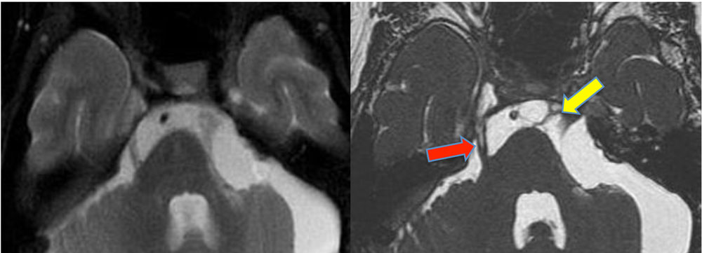
The partial volume effect/artifact is a concept so well recognized in cross-sectional imaging that it borders on being too simplistic to include in this Q&A website. At the same time, however, it is so important that I have chosen to provide a brief explanation and example for the benefit of non-radiologists who may be reviewing topics here.
The idea is easily appreciated in the example below. Here the red and yellow circles represent nerves and scar tissue residing in cerebrospinal fluid (blue). Their respective MR images appear below. The left diagram and image are 5-mm thick; the right diagram and image are 1-mm thick. The 5-mm thick voxels are of lower resolution reflecting signals that are a mixture of nerves/scar and CSF. The thin sections on the right have much less admixing of signals; the individual tissues are now sharply defined.
The idea is easily appreciated in the example below. Here the red and yellow circles represent nerves and scar tissue residing in cerebrospinal fluid (blue). Their respective MR images appear below. The left diagram and image are 5-mm thick; the right diagram and image are 1-mm thick. The 5-mm thick voxels are of lower resolution reflecting signals that are a mixture of nerves/scar and CSF. The thin sections on the right have much less admixing of signals; the individual tissues are now sharply defined.
In mathematical terms, consider a voxel that contains fractional amounts fA and fB of two materials, A and B. The MR signal from the entire voxel (SV) will then reflect the weighted average of signals SA and SB from the two components
SV = fASA + fBSB
Imperfect RF-pulse profiles may also cause to partial volume effects by exciting tissues outside the desired slice. When multiple slices are placed side, this interference is known as cross-talk.
The main strategy for decreasing partial volume artifacts is to use smaller, more sharply-defined voxels. This means thinner sections, smaller fields-of-view, and/or higher imaging matrix sizes. Using longer duration RF-pulses (or at least avoiding "fast-RF" pulse options) will improve slice profiles and create more precisely defined voxels with less excitation of adjacent tissues.
Advanced Discussion (show/hide)»
No supplementary material yet. Check back soon!
References
Kneeland JB, Shimakawa A, Wehrli FW. Effect of intersection spacing on MR image contrast and study time. Radiology 1986; 158:819-822.
Simmons A, Tofts PS, Barker GJ, Arridge SR. Sources of intensity nonuniformity in spin echo images at 1.5T. Magn Reson Med 1994: 32:121-128.
Kneeland JB, Shimakawa A, Wehrli FW. Effect of intersection spacing on MR image contrast and study time. Radiology 1986; 158:819-822.
Simmons A, Tofts PS, Barker GJ, Arridge SR. Sources of intensity nonuniformity in spin echo images at 1.5T. Magn Reson Med 1994: 32:121-128.


