|
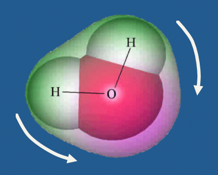
Chemical shift refers to small changes in resonant frequency due to different molecular environments of nuclei. The ¹H protons of fat, for example, are nestled within long-chain triglycerides and covered by electron clouds. These clouds partially shield the fat protons from the full effects of an externally applied magnetic field. The ¹H protons of water, however, are less shielded because their electron clouds are pulled away from them by the highly electronegative oxygen atom.
|
Because of this differential shielding by electron clouds, a fat proton experiences a slightly weaker local magnetic field and will resonate at a slightly lower frequency than a nearby water proton. The difference in frequencies is extremely small, on the order of 3.4 parts per million (ppm) or 3.4 x 10⁻⁶. At 1.5 T this corresponds to an absolute frequency difference of approximately 215 Hz. At 3.0 T the difference is twice as large, or about 430 Hz.
Consequently, if the system frequency is set to water, the signal from the lipid protons will appear to have arisen from water protons from another voxel in a lower part of the field. When image intensities are assigned in the final image, therefore, the location of fat protons will be spatially mismapped toward the lower part of the readout gradient field.
This mismapping of fat and water pixels results in artifactual white or dark bands, one-to-several pixels in width, on either side of an anatomic object. The artifact is most commonly seen around water-containing structures (liver, kidneys, optic nerves, thecal sac, muscles, nerve fascicles) surrounded by fat. Note in the image below a black band is seen on the left side of each kidney and a white band on the right. The side on which the black band appears depends on whether the the frequency-encoding gradient is increasing or decreasing from left-to-right across the image. In the kidney, knee, and leg examples shown, frequency increases from left-to-right. The image of the thecal sac is an exception; it has frequency decreasing from left-to-right. Hence the dark chemical shift band appears to the left of the thecal sac.
A very subtle form of chemical shift occurs at the edges of bones where fatty marrow abuts a water-containing joint capsule or intervertebral disk artifactually altering the apparent cortical thickness.
The size of the chemical shift artifact can be readily computed in advance based on two parameters selected prior to imaging: receiver bandwidth and size of the frequency-encode matrix. For example, if the total receiver bandwidth selected is 32 kHz and with 256 pixels in the frequency-encode direction, the bandwidth per pixel is 32,000/256 = 125 Hz. Since the fat-water frequency difference at 1.5 T is about 215 Hz, the size of the chemical-shift artifact will be (215 Hz) ÷ (125 Hz/pixel) = 1.7 pixels.
Reducing the bandwidth per pixel will accentuate this artifact. Narrow bandwidth techniques should therefore generally be avoided in locations where chemical-shift artifacts may obscure important interfaces (for instance, between the optic nerve and orbital fat).
Because the chemical-shift artifact is a spatial mismapping of MR signal based on frequency, it will typically be seen in the frequency-encode direction. (An exception occurs in echo-planar imaging, where chemical shift artifacts are commonly seen in the phase-encode direction. See the next Q&A).
Additionally, in 2DFT imaging, recall that slice selection is defined by variations in frequency. In this method of acquisition, chemical shift artifacts may occur not only within a plane of imaging, but also between slices (i.e., in the slice-select direction). Inter-slice chemical-shift artifacts may appear as dark or light "halos" around certain anatomic structures; others are less obvious and result in subtle degradation of image quality.
|
Finally, one should be aware that chemical-shift artifacts occur not only between water and fat, but at the interface of any two substances having different chemical shifts. For example, the chemical shift between silicone oil (used in ophthalmology) and water is ~4.4 ppm; between silicone gel (in breast prostheses) and water is ~4.7 ppm. Thus chemical shift artifacts may be seen in locations like the breast, head and neck, and extremities where silicone injections or implants are commonly used.
|
Advanced Discussion (show/hide)»
No supplementary material yet. Check back soon.
References
Babcock EE, Brateman L, Weinreb JC et al. Edge artifacts in MR images: chemical shift effect. J Comput Assist Tomogr 1985; 9:252-257. (One of the first descriptions of this artifact)
Birkefeld AB, Bertermann R, Eckert H, Pfleiderer B. Liquid- and solid-state high-resolution NMR methods for the investigation of aging processes of silicone breast implants. Biomaterials 2003; 24:35-46.
Dwyer AJ, Knop RH, Hoult DI. Frequency shift artifacts in MR imaging. J Comput Assist Tomogr 1985;9:16-18.
Hood MN, Ho VB, Smirniotopoulos JG, Szumowski J. Chemical shift: the artifact and clinical tool revisited. Radiographics 1999; 19:357-371 (Excellent review).
Mathews VP, Elster AD, Barker PB, et al. Intraocular silicone oil: in vitro and in vivo MR and CT characteristics. AJNR Am J Neuroradiol 1994; 15:343-7.
Smith RC, Lange RC, McCarthy SM. Chemical shift artifact: dependence on shape and orientation of the lipid-water interface. Radiology 1991; 181:225-229.
Soila KP, Viamonte M, Starewicz PM. Chemical shift misregistration effect in magnetic resonance imaging. Radiology 1984; 153:819-820. (First published description of the chemical shift artifact).


