|
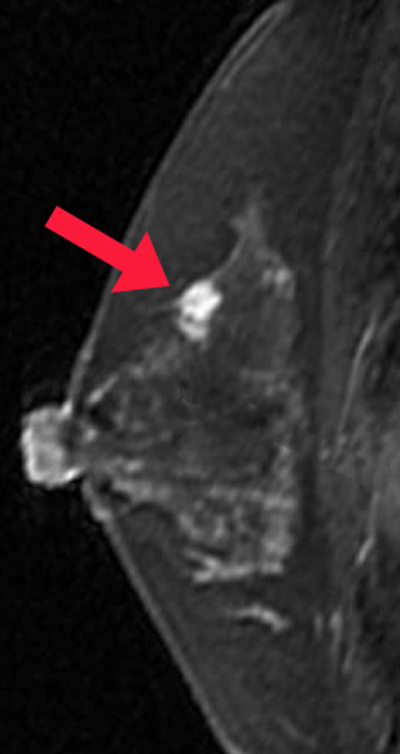
DCE breast imaging faces several challenges not present to the same extent in other parts of the body: 1) requirement to detect very small lesions in a (potentially) large volume of tissue; 2) measurement of contrast enhancement within a background of T1-bright fat; and 3) noise from respiratory and cardiac motion.
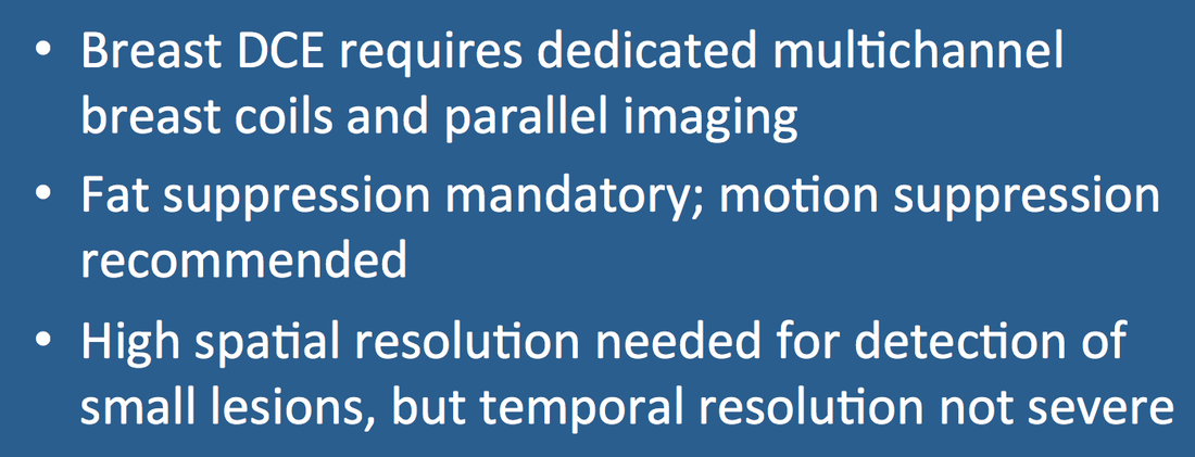
The major MR manufacturers have developed breast-optimized 3D T1-weighted gradient echo sequences known under different acronyms. These sequences all include some type of fat suppression. Typical imaging parameters are TR = 4-6 ms, TE = 1-3 ms, and flip angles = 10º-15º. Dedicated multichannel breast coils with parallel imaging must be used.
|
As with all DCE studies, a trade-off exists between spatial and temporal resolution. Due to the small size of potential tumors, the minimum spatial resolution requirements for breast DCE are an in-plane resolution of ≤ 1 mm² and a slice thickness ≤ 3 mm with no gaps. At present, DCE analysis of breast tumors is primarily observational and semi-quantitative. Temporal resolution between phases of 1½−2 minutes is therefore acceptable.
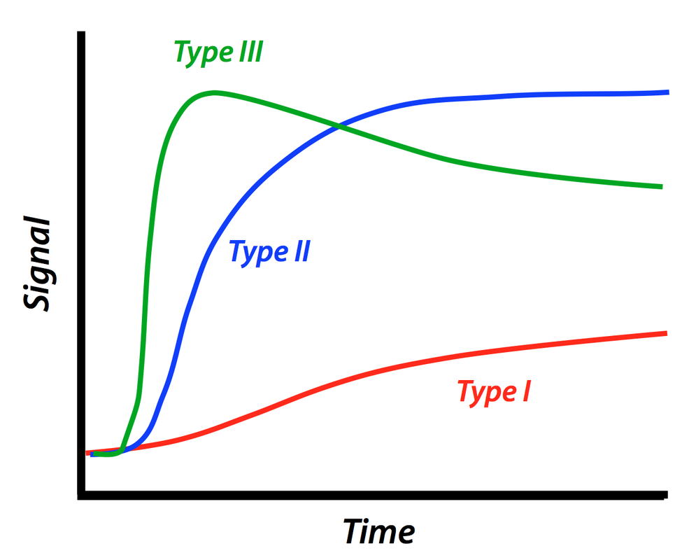
Both patterns of contrast uptake and estimated wash-in/wash-out rates from a lesion can provide clues as to its nature. A popular classification scheme by Kuhl et al. based on the shape of the contrast-time intensity curves is illustrated above. Breast masses with progressive uptake of contrast (Type I pattern) are more likely to be benign, whereas those with rapid uptake and wash-out (Type III) are more likely to be malignant. Lesions demonstrating plateau enhancement (Type II pattern) are intermediate. This classification scheme has also been applied to tumors in other organs, including the liver and prostate.
References
American College of Radiology. Breast Magnetic Resonance Imaging (MRI) accreditation program requirements, Revised 7/3/15, available at www.acr.org.
Kuhl C, Schild HH, Morakkabati N. Dynamic bilateral contrast-enhanced imaging of the breast: trade-off between spatial and temporal resolution. Radiology 2005; 236:789-800
Miyazaki M, Wheaton A, Kitane S. Enhanced fat suppression technique for breast imaging. J Magn Reson Imaging 2013; 38:981-986.
Related Questions
Our scanner offers an option called "Water Excitation". Is this just another name for Fat-Sat? What is the best technique for "first- pass" perfusion imaging?
Our scanner offers an option called "Water Excitation". Is this just another name for Fat-Sat? What is the best technique for "first- pass" perfusion imaging?


