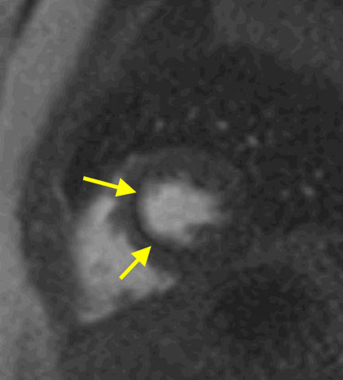
The "dark rim" artifact refers to a band of transient low signal in the endocardium during the early phases of a myocardial perfusion study. As pictured below, it is seen during the time when gadolinium first enters the left ventricle. The phenomenon is temporary, not being present after myocardial enhancement has occurred. While easily recognized by radiologists and cardiologists interpreting these studies, the transient dark rim could affect the results of automated or semiautomated measurements such as those used for up-slope analysis or parameter modeling.
The cause of the dark rim artifact is not fully established, but now believed primarily to be a manifestation of motion artifact superimposed on the Gibbs (or truncation) phenomenon. As described in a separate Q&A, the Gibbs phenomenon is seen at high contrast interfaces, such as between the brain and spinal cord (or in this case, between the myocardium and gadolinium-containing LV blood). It occurs primarily in the phase-encode direction due to sampling an inadequate number of spatial frequencies in the Fourier representation. Secondary contributing factors to the artifact may include susceptibility effect, motion, and intravoxel phase cancellations.
The dark rim artifact may be reduced by the following technical choices: 1) minimize Gibbs effect by improving image resolution via increasing number of phase-encoding lines and using parallel imaging; 2) minimize susceptibility effects by reducing contrast dose and avoiding bSSFP sequences; and 3) minimize motion artifacts via sequential k-space ordering and parallel imaging.
The more general question commonly arises: How do you know you are seeing a true perfusion defect during a first-pass study and not some sort of artifact? The answer is "experience", but the following criteria may be helpful as a starting point. A true perfusion defect typically has the following characteristics:
- It is present in at least 3-4 frames during peak passage of the Gd bolus
- It has consistent size from frame to frame
- It corresponds to a known coronary artery distribution
Conversely, artifacts such as the "dark rim" do not meet these criteria.
Advanced Discussion (show/hide)»
No supplementary material yet. Check back soon!
References
Di Bella EVR, Parker DL, Sinusas AJ. On the dark rim artifact in dynamic contrast-enhanced MRI myocardial perfusion studies. Magn Reson Med 2005; 54:1295-1299.
Ferreira PF, Gatehouse PD, Mohiaddin RH, Firmin DN. Cardiovascular magnetic resonance artefacts. J Cardiovasc Magn Reson 2013; 15:41 (Great review of all types of CMR artifacts)
Meloni A, Al-Saadi N, Torheim G, et al. Myocardial first-pass perfusion: influence of spatial resolution and heart rate on the dark rim artifact. Magn Reson Med 2011; 66:1731-8.
Saremi F, Grizzard JD, Kim RJ. Optimizing cardiac MR imaging: practical remedies for artifacts. Radiographics 2008; 28:1161-1187.
Related Questions
What is a Gibbs artifact?
What is a Gibbs artifact?


