|
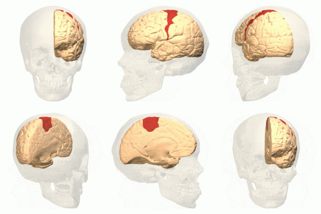
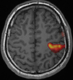
Motor cortex mapping is the among the easiest and most highly reliable of the BOLD/fMRI methods. The primary motor cortex lies along the the precentral gyrus of the frontal lobe, bordering the central sulcus (Rolandic fissure). It is immediately adjacent to and functionally related to primary somatosensory cortex, occupying the the postcentral gyrus of the anterior parietal lobe. Surgery in and around this region carries a high risk of paralysis and loss of function. Hence motor cortex mapping is the most widely used preoperative application of fMRI today.
|
 Cortical sensory homunculus
Advanced Discussion (show/hide)»
Patients with strokes, tumors, and demyelinating lesions near the motor cortex may be too weak to perform even a simple motor tasks like finger tapping. Likewise, some patients with altered mental status may not be weak but are unable to follow instructions for cued movements of the hand, face, or feet.
In these cases the motor paradigm can be modified to accommodate each patient's specific limitation. For example, if the hand is only partially dysfunctional, the finger-tapping paradigm may be converted to whole-fist clenching or squeezing of a rubber ball. For patients with complete paralysis pure sensory stimulation of the hand (by stroking or brushing) may elicit fMRI activation of both the somatosensory and primary motor cortices. Even imagined movement in some patients with total paralysis may provoke a weak BOLD-fMRI response along the sensorimotor cortices as well. Finally, resting state fMRI may allow detection of the motor cortices without any exogenous stimulus or forced movement.
American Society of Functional Neuroradiology (ASFNR). Functional Imaging Paradigms (2007). Available from http://www.asfnr.org/wp-content/uploads/ASFNR-BOLD-Paradigms.pdf (motor paradigms on pages 3-13)
Bizzi A, Biasi V, Falini A, et al. Presurgical functional MR imaging of language and motor functions: validation with intraoperative electrocortical mapping. Radiology 2008; 248:579-89.
Connelly A, Jackson GD, Frackowiak RSJ, et al. Functional mapping of activated human primary cortex with a clinical MR imaging system. Radiology 1993; 188:125-130.
Drobyshevsky A, Baumann SB, Schneider W. A rapid fMRI task battery for mapping of visual, motor, cognitive and emotional function. NeuroImage 2006; 31:732-744. (A practical and fairly comprehensive protocol for eloquent cortex mapping that can be performed in 30 min)
Hill VB, Cankurtaran CZ, Liu BP, et al. A practical review of functional MRI anatomy of the language and motor systems. Am J Neuroradiol AJNR 2019; 40:1083-1090.
Jack CR Jr, Thompson RM, Butts RK, et al. Sensory motor cortex: correlation of presurgical mapping with functional MR imaging and invasive cortical mapping. Radiology 1994;190:85–92.
Johansen-Berg H. Anatomy of the sensorimotor system. Available from Nuffield Department of Clinical Neurosciences, Oxford University at this link. (accessed Feb 2016). (It's a lot more complicated than you might think!)
Motor Cortex. Wikipedia, The Free Encyclopedia (accessed Feb 2016).
Tieleman A, Deblaere K, Van Roost D, et al. Preopertive fMRI in tumour surgery. Eur Radiol 2009; 19:2523-2534.
Ulmer JL, Klein AP, Mark LP, et al. Functional and dysfunctional sensorimotor anatomy and imaging. Semin Ultrasound CT MRI 2015; 36:220-233.
How are those activation "blobs" on an fMRI image created, and what exactly do they represent?


