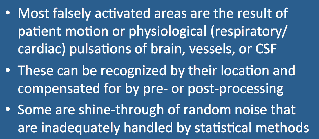
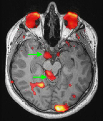
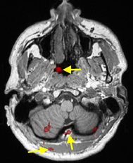
Take a look at the images below from two fMRI studies recently performed at our institution using standard pre-processing and GLM analysis. See anything wrong? How about those red blobs of "activation" that aren't within the brain? And if the images hadn't been cropped using an oval mask, many more extraneous blobs would have also been noted in "outer space" surrounding the head. We would have hoped such false positive activations would have been minimized by the rigorous statistical methods employed, but they were not.
|
Most of the blobs in the left image are due to eye motion, partial volume effects, and physiological pulsations that have not been adequately compensated/corrected. Strategies for reducing physiological noise have been described in a prior Q&A and include data scrubbing, motion correction, temporal filtering, and incorporation of measured heart and respiratory patterns as covariates in the statistical model.
|
No obvious physiological or technical source can explain the blobs in the right image, which are more likely manifestations of random noise and statistical issues, potentially related to thresholding, clustering and improper estimation of p-values. Implication of these issues forms the principal subject of a later Q&A.
Advanced Discussion (show/hide)»
No supplementary material yet. Check back soon!
References
Birn RM, Diamond JB, Smith MA, Bandettini PA. Separating respiratory-variation-related fluctuations from neuronal-acitivity-related fluctuations in fMRI. NeuroImage 2006; 31:1536-48.
Bright MG, Murphy K. Removing motion and physiological artifacts from intrinsic BOLD fluctuations using short echo data. NeuroImage 2013; 64:526-537. (Paper that focuses on resting-state and functional connectivity studies that are perhaps more affected by motion and physiological noise than task-based fMRI)
Chang C, Glover GH. Relationship between respiration, end-tidal CO2, ad BOLD signals in resting-state fMRI. NeuroImage 2009; 47:1381–1393.
Glover G, Li T, Ress,D. Image based method for retrospective correction of physiological motion effects in fMRI: RETROICOR. Magn Reson Med 2000; 44:162–167.
Nichols TE. Multiple testing corrections, nonparametric methods, and random field theory. NeuroImage 2012; 62:811-815.
Nichols T, Hayasaka S. Controlling the familywise error rate in functional neuroimaging: a comparative review. Stat Methods Med Res 2003; 12:419-446.
Related Questions
How are those activation "blobs" on an fMRI image created, and what exactly do they represent?
What are the steps for preprocessing fMRI data?
Dr. Elster, I understand you are somewhat skeptical about certain aspects of fMRI. Can you explain?
How are those activation "blobs" on an fMRI image created, and what exactly do they represent?
What are the steps for preprocessing fMRI data?
Dr. Elster, I understand you are somewhat skeptical about certain aspects of fMRI. Can you explain?


