Note: In this Q&A we adopt the convention that substances "bright" on T1-weighed images have short T1 values. This is true for most (but not all) pulse sequences. For a detailed explanation, click here.
As a rule, densely calcified tissue has a very low water content and therefore generates a very low signal on both T1 and T2 weighted images. Therefore, materials such as cortical bone and globular calcifications are routinely dark/dark on MRI.
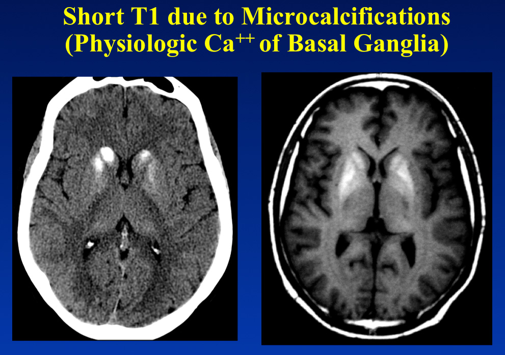
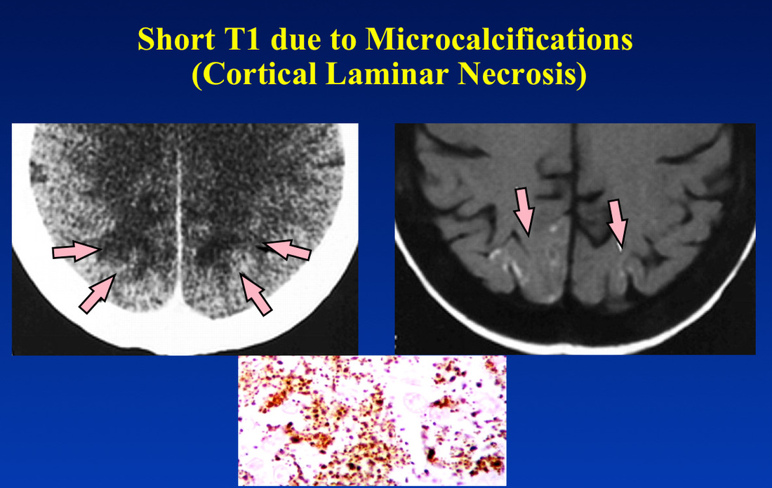
Areas containing microcalcifications, however, may sometimes appear paradoxically bright on T1-weighted images. Two common examples are shown below.
Areas containing microcalcifications, however, may sometimes appear paradoxically bright on T1-weighted images. Two common examples are shown below.
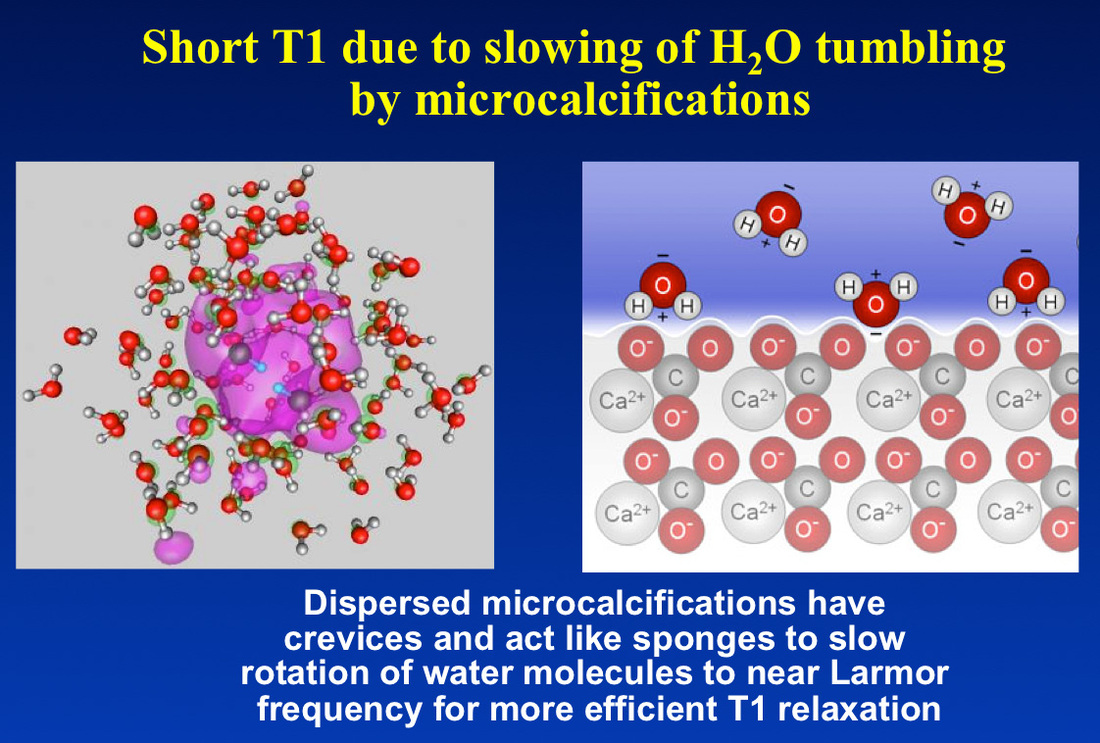
The high signal is not coming from the calcium itself, as Ca is an even-numbered element with zero spin and no intrinsic MR signal. The signal is coming from water protons, whose molecular rotation rates have been slowed to near the Larmor frequency by surface interactions with the calcium salts. This rotational slowing results in short T1 and hence brightness on T1-weighted images.
Advanced Discussion (show/hide)»
For the interested scholar, there is actually an amazing amount of literature in petrophysics concerning NMR relaxation measurements in clays and various porous rocks. This is important in mining and drilling for oil and gas. The relaxation effects involve restricted diffusion as well as T1/T2 shortening, and is a function of pore size and a number of other factors. This PhD thesis link provides a good review and starting point for further investigation.
Related Questions
What is T1 relaxation?
Can you explain a little more about the dipole-dipole interaction? I still don't quite understand.
What is T1 relaxation?
Can you explain a little more about the dipole-dipole interaction? I still don't quite understand.


