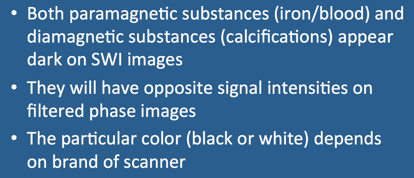
In early iterations of SWI in the 1980's and 1990's, phase information was largely discarded in favor of magnitude data to produce "black-blood" MR venograms and BOLD (Blood Oxygen Level Dependent) functional MRI maps. By the mid-1990s, however, it was recognized that phase information could be used to distinguish paramagnetic substances (like iron and blood) from diamagnetic substances (like calcifications). Modern SWI sequences produce filtered phase images where (paramagnetic) blood products have signal intensities opposite to (diamagnetic) calcifications. Some care must be taken to determine which is which, however.
|
To establish a reference, blood in cortical veins or dural sinuses may be used to determine the expected "color" (i.e., black or white) of paramagnetic substances in the phase image. Alternatively, areas of known calcification (e.g., pineal gland, choroid plexus, or dorsum sellae) may be used as a reference for diamagnetic substances.
An important caveat is that the "colors" of blood and calcium on SWI phase images are scanner- dependent. Siemens and Canon use so-called "left-handed" reference schemes where blood products appear bright; GE and Philips use a "right-handed" reference where blood products appear dark. |
Advanced Discussion (show/hide)»
No supplementary material yet. Check back soon!
References
Barbosa JHO, Santos AC, Salmon CEG. Susceptibility weighted imaging: differentiating between calcification and hemosiderin. Radiol Bras 2015; 48:93-100.
Duyn J. MR susceptibility imaging. J Magn Reson 2013; 229:198-207. (good historical review)
Haacke EM, Xu Y, Cheng Y-C, Reichenbach JR. Susceptibility weighted imaging (SWI). Magn Reson Med 2004; 52:612-618. (Describes processing method now used for most commercial SWI sequences)
Haacke EM, Mittal S, Wu Z, Neelavalli J, Cheng Y-CN. Susceptibility-weighted imaging: technical aspects and clinical applications, part 1. AJNR Am J Neuroradiol 2009; 30:19-30. (technical features)
Mittal S, Wu Z, Neelavalli J, Haacke EM. Susceptibility-weighted imaging: technical aspects and clinical applications, part 2. AJNR Am J Neuroradiol 2009; 30:232-252. (clinical applications)
Yamada N, Imakita S, Sakuma T, Takamiya M. Intracranial calcification on gradient-echo phase image: depiction of diamagnetic susceptibility. Radiology 1996; 198:171-178. (first use of phase mapping to distinguish diamagnetic from paramagnetic substances on SWI)
Related Questions
How are susceptibility-weighted images obtained and processed?
How are susceptibility-weighted images obtained and processed?

