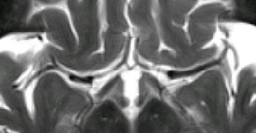
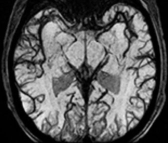
 Vertebral artery dissection (arrow).
Note dark blood
 "flow voids" in other arteries (but not the veins).
Dark Blood MR Angiography is vascular imaging strategy wherein the signal from flowing blood is suppressed (rendering it "black") rather than enhanced as it is in conventional Bright Blood MRA techniques. Rapidly flowing or turbulent blood has a naturally low signal because of phase-dispersion and time-of-flight signal losses. These effects may be further accentuated by application of flow-spoiling gradients, saturation bands, and/or inversion pulses. The well-recognized vascular "flow voids" seen on routine MR imaging represent a crude form of dark-blood MRA.
In theory, dark-blood MRA has some unique advantages compared to bright-blood techniques. Flow separation and turbulence, which cause unwanted signal losses and overestimation of stenoses on bright-blood MRA, paradoxically improve image quality on dark-blood MRA. Because intraluminal signal is suppressed, vessels containing "black blood" do not generate pulsation (ghosting) artifacts. Additionally, the lack of intraluminal signal allows the walls of vessels (or cardiac chambers) to be clearly delineated. For these reasons, dark-blood techniques are predominantly used in cardiac imaging and for evaluation of diseases of the vessel wall (atherosclerotic plaque and dissection).
In theory, dark-blood MRA has some unique advantages compared to bright-blood techniques. Flow separation and turbulence, which cause unwanted signal losses and overestimation of stenoses on bright-blood MRA, paradoxically improve image quality on dark-blood MRA. Because intraluminal signal is suppressed, vessels containing "black blood" do not generate pulsation (ghosting) artifacts. Additionally, the lack of intraluminal signal allows the walls of vessels (or cardiac chambers) to be clearly delineated. For these reasons, dark-blood techniques are predominantly used in cardiac imaging and for evaluation of diseases of the vessel wall (atherosclerotic plaque and dissection).
Notwithstanding these specific cardiovascular applications, dark-blood MRA has never taken hold as a widely used method for the head, abdomen, or extremities. Although an excellent method for imaging high-flow vessels, dark-blood techniques are less sensitive to slower flow states. "Black" blood is difficult to separate from adjacent low-signal non-vascular structures, such as calcifications, cortical bone, and air.
To convert a set of dark-blood source images into an angiographic-style picture requires a minimum intensity projection method. By comparison, bright-blood MRA is typically displayed using a maximum intensity projection algorithm. The minimum intensity method required for dark-blood MRA requires considerable manual editing to exclude air (both inside and around the patient) as well as cortical bone and other low signal non-vascular structures. As such, dark-blood MRA studies are usually displayed as tomographic sections rather than as angiogram-like projections.
A dark-blood MR angiogram is not the same as simply inverting the gray-scale image of a bright-blood MR angiogram. Completely different methods are responsible for the vascular contrast observed.
|
Thin-slice 2D or 3D Fast Spin Echo (FSE) sequences can be used to generate dark-blood MRA's that can be reconstructed using multiplanar or curvilinear displays. This method is especially useful in the brain where a T2-weighted FSE sequence produces high signal from CSF in contrast to the black vessels.
|
|
The degree of vascular dephasing may be increased by applying weak, diffusion-like spoiling gradients along all three axes in a 3D GRE sequence. The result is an increased sensitivity toward more slowly flowing blood in smaller vessels. This method is part of Canon's V-TRACE ("Variable True Rate Angiography with Combined Encodings") technique. V-TRACE acquires acquires a bright-blood MRA image simultaneously without flow spoiling at a shorter TE. The black blood image is usually not displayed separately but subtracted from the bright-blood image to improve visualization of smaller vessels.
|
The above-described methods of black-blood imaging rely primarily upon MR flow phenomena to create dark intraluminal signal (potentially supplemented by flow-spoiling gradients or saturation bands). Two additional important methods of dark-blood imaging, Inversion Recovery (IR) Black Blood MRA and Susceptibility-Weighted (SW) Black Blood MRA, exploit the relaxation times of blood to create a black blood effect. These important methods are described in detail in subsequent Q&A's.
Advanced Discussion (show/hide)»
No supplementary material yet. Check back soon.
References
Edelman RR, Mattle HP, Wallner B et al. Extracranial carotid arteries: evaluation with "black blood" MR angiography. Radiology 1990; 177:45-50.
Edelman RR, Chien D, Kim D. Fast selective black blood MR imaging. Radiology 1991; 181:655-660.
Kimura T, Ikedo M, Takemoto S. Hybrid of opposite-contrast MR angiography (HOP-MRA) combining time-of-flight and flow-sensitive black-blood contrasts. Magn Reson Med 2009; 62:450-458. (This is the technical description underlying Canon's V-TRACE method).
Related Questions
How do you create an MR angiogram?
How do you create an MR angiogram?


