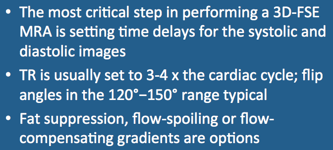
To set up a 3D-FSE MRA scan, the following steps are typically performed:
|
1. Reliable EKG or peripheral pulse gating is first established.
2. A low-resolution localizer is obtained to identify anatomy and position a 3D FSE slab over area of interest. Depending on the area scanned, a spatial saturation pulse may needed to reduce the size of the 3D slab and prevent wrap-around. 3. Proper delay times for systolic and diastolic imaging must next be determined. For more proximal vessels technologists may be able to estimate these values based on experience. For more distal vessels a calibration procedure is generally advised. This involves running an additional cine scout sequence using single shot fast spin echo (SSFSE/HASTE) technique. Placing a cursor over the target vessel from these cine scout images, maximum and minimum signal intensities can be graphically displayed and optimal systolic and diastolic delay times computed. Note that these times may differ significantly between the two extremities as a result of atherosclerosis or variant anatomy. 4. Once the systolic and diastolic delay times have been determined, the final scan may be run. Typically the TR value is set to about 3-4 times the cardiac (R-R) interval. The flip angle (α) is typically chosen in the range of 120º−150º, but to detect smaller and more slowly flowing vessels, values closer to 90º may be used. 5. Fat suppression is not technically necessary since fat is removed by the subtraction process. However, a STIR or SPAIR pulse is sometimes used to suppress fat on the individual systolic and diastolic source images that are typically reviewed as part of the diagnostic workflow. |
6. The 3D FSE pulse sequence is most sensitive to motion when artery of interest is oriented in the direction of the readout gradient. This is the result of endogenous spoiling (disruption) of transverse coherences by the alternating echoes in the FSE train. Accentuated systolic signal loss ("flow void") may be produced by applying additional flow-spoiling gradients as an option. This improves the visibility of smaller, peripheral, and more slowly flowing vessels.
7. Alternatively, sometimes too much turbulence and flow disruption occurs in larger central arteries without significant stasis in diastole to produce a measurable intraluminal signal. In this situation, flow-compensating gradients (similar to those used for gradient-moment nulling) are employed. These gradients increase the diastolic signal so that the final subtraction image (diastolic − systolic) will provide a good representation of the high-flow artery.
7. Alternatively, sometimes too much turbulence and flow disruption occurs in larger central arteries without significant stasis in diastole to produce a measurable intraluminal signal. In this situation, flow-compensating gradients (similar to those used for gradient-moment nulling) are employed. These gradients increase the diastolic signal so that the final subtraction image (diastolic − systolic) will provide a good representation of the high-flow artery.
Advanced Discussion (show/hide)»
Similar to SPACE/CUBE/VISTA, variable flip angle gated 3D-FSE MRA techniques are now being developed and deployed by various manufacturers. These methods are also being integrated into parallel imaging to further reduce imaging times and artifacts.
References
Wheaton AJ, Miyazaki M. Non-contrast enhanced MR angiography: physical principles. J Magn Reson Imaging 2012; 36:286-304.
Wheaton AJ, Miyazaki M. Non-contrast enhanced MR angiography: physical principles. J Magn Reson Imaging 2012; 36:286-304.

