In the 1950s and 1960s considerably progress was made in explaining the the physical principles underlying T1 and T2 relaxation in pure liquids and simple solutions. It was soon recognized that these theories gave incorrect predictions when applied to biological specimens, however. Several factors accounted for these discrepancies:
- Tissues contain macromolecules (proteins, phospholipids, polysaccharides, fat) in addition to water.
- Tissues are compartmentalized.
- A small but significant portion of the water in tissue is not free, but tightly or moderately bound to macromolecules.

Hydration layers around a macromolecule
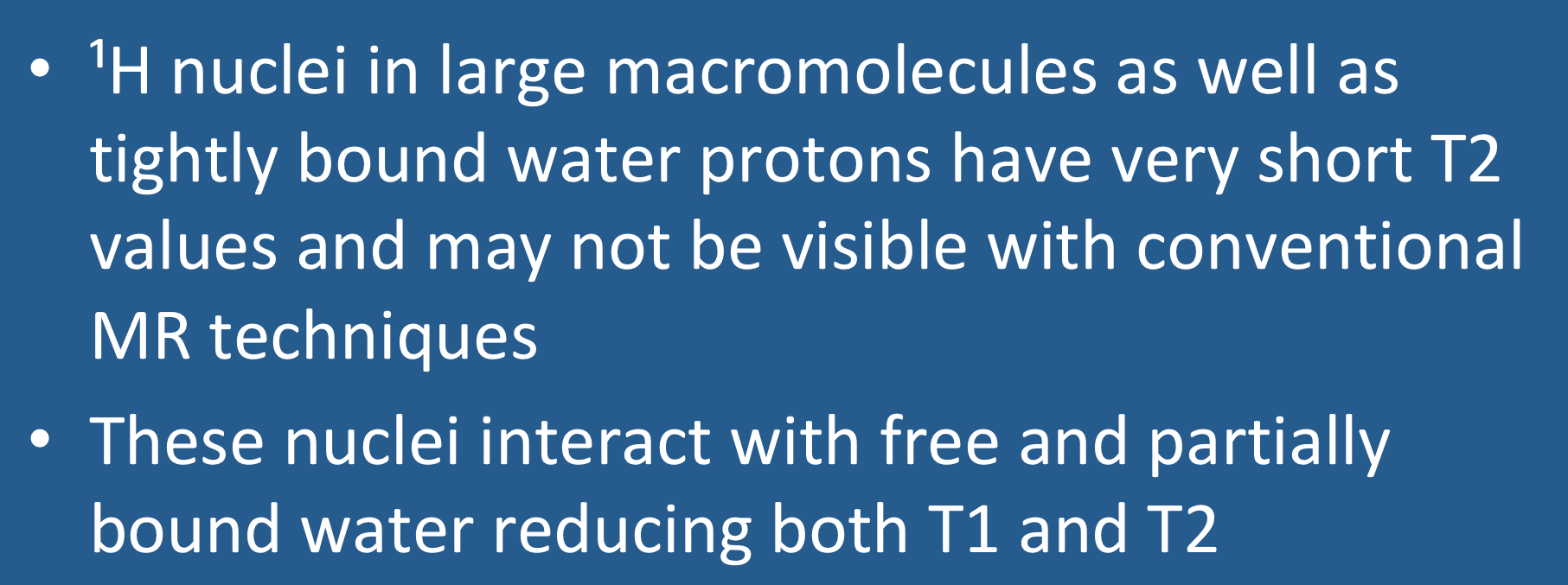
The cytosol of mammalian cells is extremely crowded, and there is restricted movement of larger proteins that are forced into globular shapes. ¹H protons deep within these macromolecules have restricted motions, resulting in very short T2 values (often 1 ms or less). The signals from these protons decay so quickly that they may be undetectable by "conventional" MR imaging methods using TE values longer than 1 ms. Tightly bound ¹H2O molecules immediately adjacent to the macromolecules or within their crevices behave in a similar fashion, possessing very short T2 values with signals decaying so rapidly that they may also be undetectable by conventional imaging techniques and part of the so-called MR "dark matter". Although not directly visualized, these bound water molecules they can undergo dipolar interactions and chemical exchange both with macromolecular protons and those in the free water pool. There is also an intermediate layer of water molecules with characteristics between the free and bound forms.
The presence of macromolecules slows the normally rapid rotation and diffusion of the smaller water molecules by means of hydrogen bonding and other interactions. Reduced motion means that a higher proportion of HOH nuclei will experience both static fields (reducing T2) and interactions near the Larmor frequency (reducing T1). The degree of T1 and T2 shortening depends on: 1) the concentration of the macromolecules, 2) their size, and 3) the number of hydrophilic groups (amine, hydroxyl, and carboxyl) to bind water molecules.
The interaction between free and bound pools is discussed more completely in several later Q&A's where the concept of magnetization transfer is explained.
The interaction between free and bound pools is discussed more completely in several later Q&A's where the concept of magnetization transfer is explained.
Advanced Discussion (show/hide)»
No supplementary material yet. Check back soon!
References
Edzes HT, Samulski ET. Cross-relaxation and spin diffusion in the proton NMR of hydrated collagen. Nature 1977;265:521-523. (A famous paper first showing how the interaction between water and proteins affects relaxation times.)
Halle B. Water in biological systems: the NMR picture. In: Bellissent-Funel M-C (ed.). Hydration processes in biology : theoretical and experimental approaches. IOS Press, Clifton, VA. 1999: 233-249.
Levy Y, Onuchic JN. Water and proteins: a love-hate relationship. PNAS 2004;101:3325-6.
Zhang L, Wang L, Kao Y-T, et al. Mapping hydration dynamics around a protein surface. PNAS 2007; 104:18461-6. (There is a lot of current research activity concerning how water molecules in the hydration sphere as well as some embedded in the interstices of macromolecules affect and regulate biological activity.)
Related Questions
Can you explain a little more about the dipole-dipole interaction? I still don't quite understand.
How can you predict the T1 and T2 values of different tissues?
What is magnetization transfer?
Why are some nuclei "invisible" on MRI?
Can you explain a little more about the dipole-dipole interaction? I still don't quite understand.
How can you predict the T1 and T2 values of different tissues?
What is magnetization transfer?
Why are some nuclei "invisible" on MRI?
