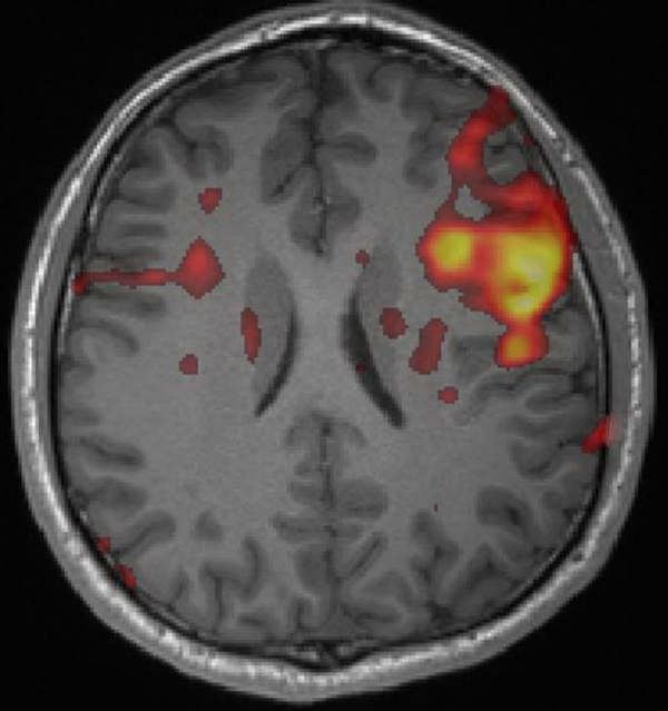
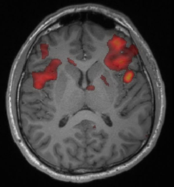
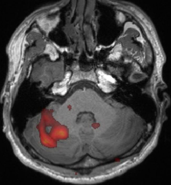
 "Classic" and "Dual Stream" models of language processing. Classic model is centered on Wernicke's (receptive) and Broca's (expressive) areas. "Dual stream" model incorporates these areas into a ventral stream (red) serving comprehension and a dorsal stream (blue) for articulation.
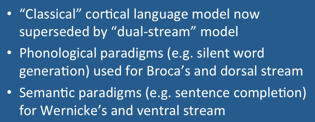
Over the last decade a more complex "dual stream" model has been embraced by much of the neuroscience community, recognizing the role of additional involvement of the temporal, parietal, and frontal lobes in language processing. In this model a bilaterally organized ventral stream processes speech signals for comprehension and a strongly left-dominant dorsal stream maps acoustic speech to articulatory networks in the frontal lobe.
bilateral Broca's areas, left greater than right (B); and right cerebellum (C)
Advanced Discussion (show/hide)»
Bilingual Patients
Bilingual subjects demonstrate few fMRI differences in their temporal lobe language association (Wernicke's) areas from single language patients. Broca's areas are different, however. Here the distinction is whether the second language was acquired early (in childhood) or late (as teenagers or adults). Late bilingual subject typically have separate expressive language areas while these areas are shared in early bilingual subjects.
Kim K et al. Distinct cortical areas associated with native and second languages. Nature 1997; 388, 171-174.
Hemispheric Dominance
The degree of hemispheric dominance in an fMRI activation experiment can be quantified by a Laterality Index (LI) defined as
LI = (L− R) / (L + R)
Using this metric, a highly left hemisphere dominant person would receive a score of +1, a highly right dominant person would receive −1, and a perfectly balanced person would receive a score of zero (0). However, the LI is somewhat arbitrary in that it may change depending on the statistical thresholds used to define activation.
Agarwal S, Sair HI, Guiar S, Pillai JJ. Language mapping with fMRI. Current standards and reproducibility. Top Magn Reson Imaging 2019; 28:225-233. [DOI Link] (excellent recent review).
Black DF, Vachha B, Mian A, et al. American Society of Functional Neuroradiology-recommended fMRI paradigm algorithms for presurgical language assessment. AJNR Am J Neuroradiol 2017; 38:E65-73. [DOI Link]
Drobyshevsky A, Baumann SB, Schneider W. A rapid fMRI task battery for mapping of visual, motor, cognitive and emotional function. NeuroImage 2006; 31:732-744. [DOI Link] (A practical and fairly comprehensive protocol for eloquent cortex mapping that can be performed in 30 min)
Hickok G. The functional neuroanatomy of language. Phys Life Rev 2009; 6:121-143. [DOI Link] (good review by the originator of the modern "dual stream" model of speech processing)
Hill VB, Cankurtaran CZ, Liu BP, et al. A practical review of functional MRI anatomy of the language and motor systems. Am J Neuroradiol AJNR 2019; 40:1083-1090. [DOI Link]
Klein AP, Sabsevits DS, Ulmer JL, Mark LP. Imaging of cortical and white matter language processing. Semin Ultrasound CT MRI 2015; 36:249-259. [DOI Link]
Price CJ. A review and synthesis of the first 20 years of PET and fMRI studies of heard speech, spoken language and reading. Neuroimage 2012; 62:816-847. [DOI Link]
Smits M, Visch-Brink E, Schraa-Tam CK, et al. Functional MR imaging of language processing: an overview of easy-to-implement paradigms for patient care and clinical research. Radiographics 2006; 26:S145-158. [DOI Link]
Zaca D, Jarsol S, Pillai JJ. Role of semantic paradigms for optimization of language mapping in clinical fMRI studies. AJNR Am J Neuroradiol 2013 34: 1966-1971. [DOI Link]
Why do you have to do an "on-off" comparison? Why not just measure the absolute BOLD signal instead?
How are those activation "blobs" on an fMRI image created, and what exactly do they represent?


