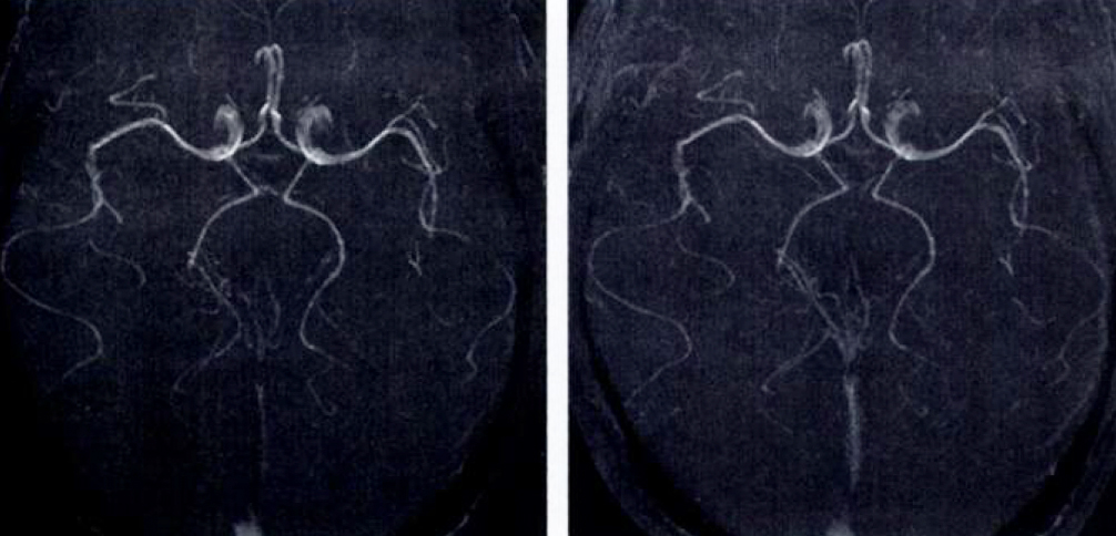
As described in a previous Q&A, magnetization transfer (MT) is the physical process by which macromolecules and their closely associated water molecules (the "bound" pool) cross-relax with protons in the free water pool. The interaction between the two pools can be modulated by applying radiofrequency (RF) energy exclusively to the bound pool using specially designed off-resonance MT pulse(s).
Some of this deposited energy is then transferred to the free water pool primarily via dipole-dipole interactions. Depending on the degree of coupling between the pools, the free water pool becomes partially saturated. If the free water pool is subsequently imaged using routine RF pulses and gradients, its signal will be reduced secondary to an MT effect. I personally find it helpful to think of MT pulses as "macromolecule sat pulses" which selectively suppress tissues with significant water-macromolecule interactions.
Some of this deposited energy is then transferred to the free water pool primarily via dipole-dipole interactions. Depending on the degree of coupling between the pools, the free water pool becomes partially saturated. If the free water pool is subsequently imaged using routine RF pulses and gradients, its signal will be reduced secondary to an MT effect. I personally find it helpful to think of MT pulses as "macromolecule sat pulses" which selectively suppress tissues with significant water-macromolecule interactions.
The suppression of background tissue by MT pulses makes the technique especially useful as an adjunct to MR angiography (MRA). Small vessel detail and overall image contrast are significantly improved in time of flight (TOF) MRA when MT pulses are employed.
Magnetization transfer techniques are equally effective for both non-contrast and contast-enhanced MRA. Because gadolinium (Gd) enhancement is caused by a water-Gd ion interaction (not macromolecular cross-relaxation), MT pulses suppress the signal from background tissues and render Gd-enhanced areas more conspicuous.
Limitations and disadvantages of MT-assisted MRA include: 1) a slight prolongation of imaging time (extra time is required to perform the MT pulses); and 2) tissue heating due to energy deposition from the MT pulses. Notwithstanding these limitations, MT pulses are widely used in clinical MRA studies, especially of the brain.
Advanced Discussion (show/hide)»
All the major vendors use the generic terms magnetization transfer, MT, or MTC to refer to this technique. Toshiba, however, uses a much longer but more descriptive trade name, SORS-STC, which stands for "Slice-Selective Off-Resonance Sinc Pulse Saturation Transfer Contrast".
References
de Boer RW. Magnetization transfer contrast. Part 1: MR Physics. Philips Medical Systems MedicaMundi 1995;40:64-73.
de Boer RW. Magnetization transfer contrast. Part 2: Clinical applications. Philips Medical Systems MedicaMundi 1995;40:74-83.
Elster AD, King JC, Mathews VP, Hamilton CA. Cranial tissues: appearance at gadolinium-enhanced and nonenhanced MR imaging with magnetization transfer contrast. Radiology 1994; 190:541-546.
Elster AD, King JC, Mathews VP, Hamilton C. Improved detection of gadolinium enhancement using magnetization transfer imaging. Neuroimaging Clin N Am 1994; 4:185-192.
Mathews VP, Elster AD, King JC et al. Combined effects of magnetization transfer and gadolinium in cranial MR imaging and MR angiography. AJR Am J Roentgenol 1995; 164:169-172.
Henkelman RM, Stanisz GJ, Graham SJ. Magnetization transfer in MRI: a review. NMR Biomed 2001;14:57-64.
Ulmer JL, Mathews VP, Hamilton CA, Elster AD, Moran PR. Magnetization transfer or spin-lock? An investigation of off-resonance saturation pulse imaging with varying frequency offsets. AJNR Am J Neuroradiol 1996; 17:805-819.
Related Questions
How does the presence of macromolecules affect T1 and T2?
What is magnetization transfer?
How is contrast generated by magnetization transfer? Also, what are MTI, MTC, and MTR?
How does the presence of macromolecules affect T1 and T2?
What is magnetization transfer?
How is contrast generated by magnetization transfer? Also, what are MTI, MTC, and MTR?

